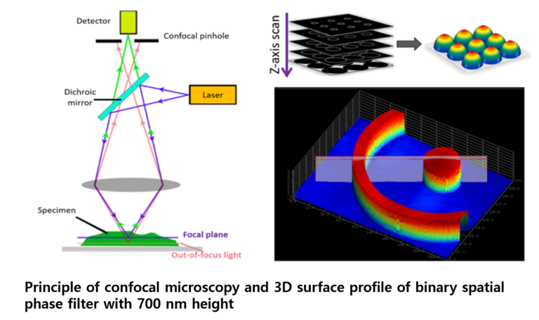
Confocal microscopy has proven to be one of the most important advances in optical microscopy. The principle of the confocal approach is to use a pinhole as a spatial filter to collect light only from the focal plane and to eliminate the reflected light from above and below the focal plane. Confocal microscopy offers several advantages over conventional widefield optical microscopy, including the ability to control depth of field, elimination or reduction of background information away from the focal plane, and the capability to collect serial optical sections from thick specimens. Since confocal microscopy can provide extremely high-quality and three-dimensional images, it is widely used in a variety of fields such as biological imaging and industrial measurement. In BOOM Lab we are developing a real-time three-dimensional confocal microscopy based on dual-detection technique and the chromatic confocal method.
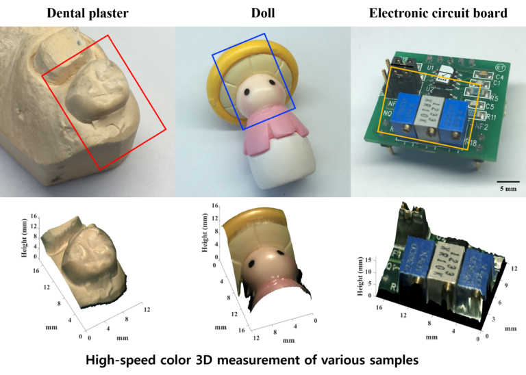
Color 3D measurement
Optical 3D measurements are widely used in a variety of industrial and biomedical applications requiring fast and accurate 3D surface profiles without touching objects. BOOM Lab has developed a high-speed color 3D reconstruction method based on direct-view confocal microscopy. By utilizing a pinhole array, the height information can be acquired from multiple pinholes in parallel. In addition, the color information of the object at focus can be obtained with a high-speed color CMOS camera with white-light illumination. The precise color 3D surface can be reconstructed by superimposing the color image over the height information. We anticipate this technology to be useful in various fields, such as biomedical imaging and industrial non-contact inspection.
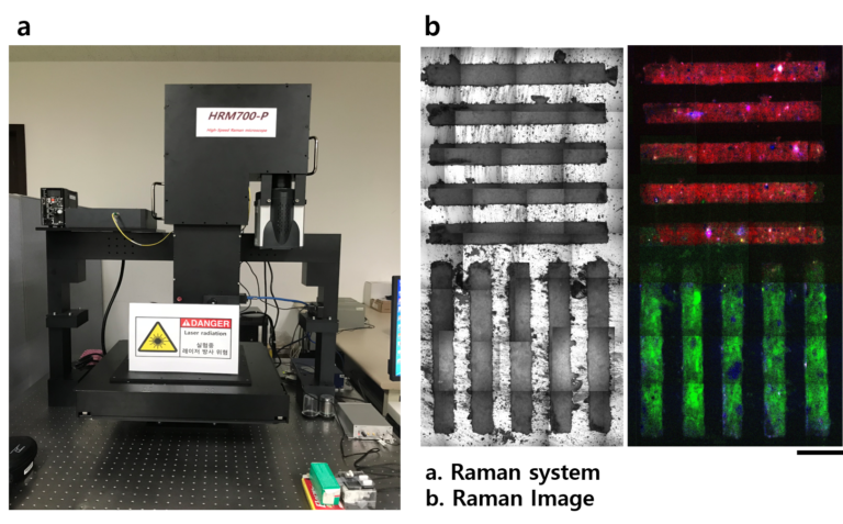
High-speed confocal Raman Microscopy
Confocal Raman microscopy is used in a variety of research fields because it allows for label-free sample investigation at a microscopic level. However, Raman imaging at high temporal and spectral resolution has been technically challenging because Raman microscopy requires a long exposure time to detect a weak Raman scattering signal. We have developed a high-speed confocal Raman system based on line illumination and the dual-mirror scan method. We anticipate this technology to be useful for high-speed large-area Raman microscopic imaging because of its high spectral rate of up to 25,600 spectra/s.