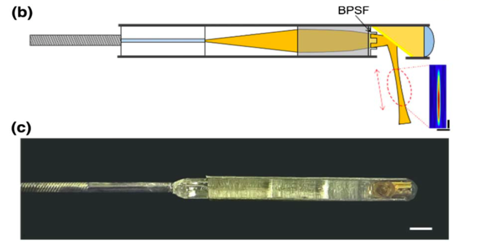
One of the greatest advantages of optical technology for biomedical imaging is that it can provide high-resolution images compared to other medical imaging devices, such as MRI, CT, and ultrasound. Endoscopic imaging catheters are emerging technologies for clinical and preclinical studies that provide high-resolution tomographic images of endoluminal organs, such as blood vessels, the esophagus, and auditory tubes. We are developing new endoscopic imaging catheter systems that offer higher spatial resolution with extended imaging depth for a variety of biomedical applications.
Optical biopsy
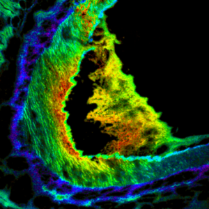
Biopsy followed by histopathology has been regarded as a gold standard for the diagnosis of cancer and many other diseases. Traditional histopathology requires numerous steps, such as tissue collection, fixation, slicing, and staining, which leads to prolonged examination time. Optical microscopy techniques are promising candidates in histopathology in that they provide the microscopic features of tissue in situ. For example, innovative optical techniques such as confocal reflection/fluorescence microscopy, multi-photon microscopy, second harmonic generation, fluorescence lifetime imaging, and Raman microscopy, enable rapid visualization of tissue samples. These optical biopsy techniques promise rapid, accurate, and safe diagnosis by providing various biochemical and morphological information on a microscopic scale.
Multimodal Microscopy
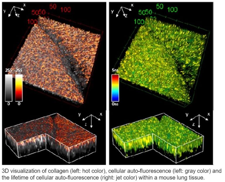
In the biomedical field, it is important to obtain both structural and functional information for the multilateral understanding of biological samples such as tissues and cells. Multimodal microscopy provides such comprehensive information by integrating various microscopy tools on one platform. For example, one of our multimodal microscope systems includes a fluorescence lifetime imaging microscopy (FLIM) to obtain biochemical information along with other microscopies to enable optical sectioning for precise structural imaging.
Cardiovascular disease
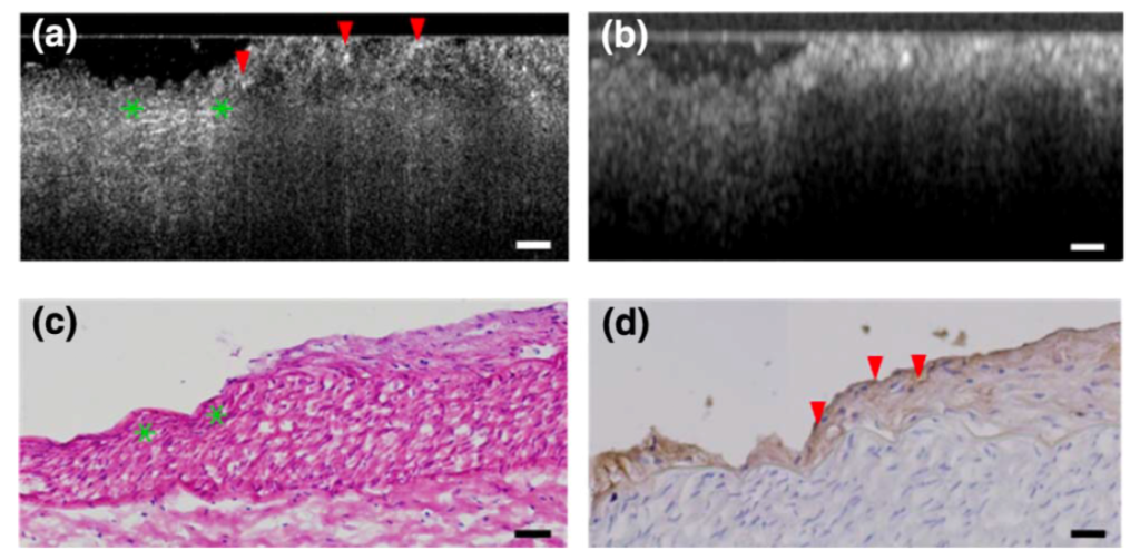
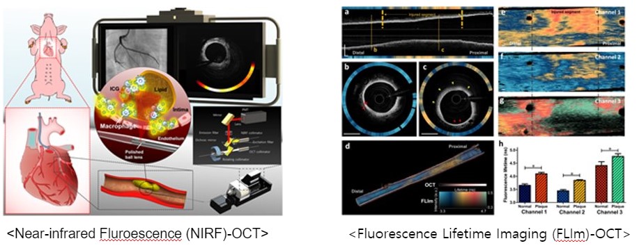
Cardiovascular disease, especially coronary artery disease such as atherosclerosis, is the leading cause of death worldwide. Progression of the atherosclerotic plaque narrows the coronary arteries, impedes blood flow to the myocardium, and increases the risk of fatal events such as myocardial infarction and heart attack. Unfortunately, current medical imaging technologies, including intravascular ultrasound and intravascular optical coherence tomography (OCT), are unable to obtain biochemical information, such as inflammation and arterial composition, which are closely related to the progression of arterial disease. Thus, in BOOM Lab, we are working to develop advanced multimodal catheter-based OCT technology to provide a comprehensive picture of atherosclerotic plaque for accurate diagnosis. We have developed multimodal intravascular OCT systems by combining OCT with near-fluorescence fluorescence (NIRF) or fluorescence lifetime imaging microscopy (FLIm). NIRF-OCT can visualize both microstructure and inflammation information in coronary arteries. FLIm-OCT provides compositional biochemical information without any contrast agents. We have recently been focusing on the development of clinically available FLIm-OCT devices and theranostic NIRF-OCT systems capable of photodynamic therapy.
Machine learning for optical image processing
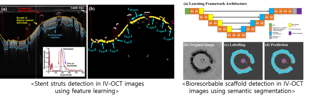
Recent advances in optical imaging techniques for biomedical applications have provided new opportunities to understand the pathology underlying various diseases. More accurate lesion evaluation is enabled based on quantitative analysis on the biomedical optical images, which requires time-consuming and labor-intensive segmentation process. Machine learning has achieved the state-of-the-art segmentation results in a variety of automated methods for segmentation of specific lesions or structures in the optical images, especially in IV-OCT images. Recently, we are interested in developing methods for segmentation and image quality enhancement (restoration) using novel deep learning techniques, such as convolutional neural networks or generative adversarial network.